Echocardiography is a highly versatile phenotyping process which makes it the non-invasive tool of choice. While other non-invasive (MRI, PET) or invasive (cardiac catheterization, implanted telemetry) techniques can provide superior data in select aspects of cardiovascular phenotyping, none has the versatility that allows rapid and comprehensive evaluation of myocardial, valvular and vascular structure and function in animal models as echocardiography does. Nevertheless, based on our experience in the Core, we strongly believe that these techniques are complementary and should be used in an integrated manner applied selectively to the specific needs of the projects.
There has been great progress made in murine echocardiography since the first reports of M-mode images. The days of using M-mode images of the left ventricle with very limited spatial resolution as the sole method for evaluating cardiac phenotypes have passed.
A comprehensive mouse echocardiogram today includes:
- two-dimensional short- and long-axis cineloops of the heart from the parasternal view
- two-dimensional guided M-mode image of the left ventricle and basal structures
- Doppler recordings of trans-mitral, aortic, and pulmonary valve velocities
- Doppler tissue recordings of mitral annular and left ventricular segmental wall motion abnormalities
Under special experimental conditions, additional image acquisition protocols can be applied (see below).
Service and data output details
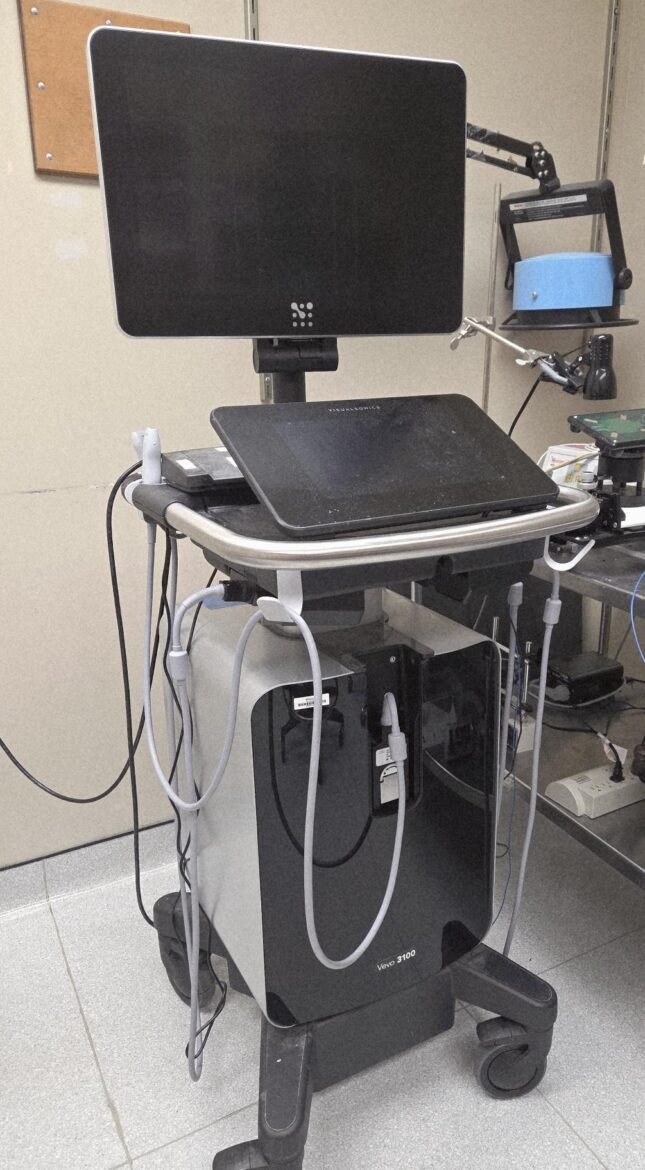
Non-invasive transthoracic cardiac ultrasound exam is performed under various anesthetic protocols (unless status of animal precludes anesthesia). A FujiFilm VisualSonic echocardiography machine with multiple transducers is used to characterize the structure and function of the heart and great vessels.
Data provided at study end: Spreadsheet with various echo parameters.
Non-invasive single transthoracic cardiac ultrasound exam performed under light general anesthesia that includes two-dimensional and M-mode images of the left ventricle.
Full echocardiograms to document the outcome of a specific surgeries (such as TAC, MI, I/R). This echo may include two-dimensional and M-mode images and analysis of the left ventricle, or Doppler recordings of trans-valvular flow indices (gradients), or segmental wall motion abnormalities..
Echo to delineate structural and functional abnormalities of the aorta or other vessels. Includes an analysis of these parameters and data reporting.
This echo is charged on a “per mother” basis. Echo includes delineating the structure and function of hearts of embryos. This is a terminal study in which mother and pups will be euthanized at study end. Echos will be performed on embryos in utero whereby the uterus has been externalized while the mother is under gas anesthesia.
Includes studies that the echocardiographer and PI have discussed in detail and that are not offered on a routine basis. Analysis and data reporting will be included.
Previously unscheduled echoes that are necessary to do immediately because mouse’s clinical condition.
Echocardiograms of any species other than mice.